X-Ray Cervical Spine AP & Lat
Comprehensive Radiographic Examination of the Neck
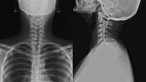
Overview of the X-Ray Cervical Spine AP and Lat
The X ray cervical spine AP lat is a comprehensive radiographic examination that visualises the cervical spine, which includes the first seven vertebrae (C1-C7). This cervical spine X ray is crucial for identifying and evaluating various conditions affecting the bones, joints, and soft tissues of the neck. The AP (anteroposterior) view involves taking an X-ray from the front to the back of the patient, while the lat (lateral) view captures the spine from the side. Together, these views provide a thorough assessment of the cervical spine, helping doctors diagnose a range of conditions, plan treatments, and monitor the progression of diseases or the healing process after surgeries.
Cost of X-Ray Cervical Spine AP and Lat
The X ray cervical spine AP lat cost can vary depending on factors such as the location, type of diagnostic centre, and the city where the test is conducted. Generally, the X ray cervical spine AP lat cost for this test falls between ₹500 to ₹2,000 or more. Government hospitals tend to offer the most affordable rates, while private clinics and advanced diagnostic centres may charge higher fees.
Use of X-Ray Cervical Spine AP and Lat
The X ray cervical spine AP lat is used to diagnose and evaluate a wide range of conditions affecting the cervical spine. Some of the most common reasons for recommending this test include:
What is the Procedure for X-Ray Cervical Spine AP and Lat
The procedure for a cervical spine X ray involves several steps to ensure accurate imaging of the cervical spine. The process typically includes:
- Preparation: Patients are asked to remove any metal objects, such as jewellery, glasses, or clothing, that could interfere with the cervical spine X ray images. They should also inform the doctor if they are pregnant or suspect pregnancy.
- Positioning for AP View: The patient stands erect, unless in cases of trauma, where they may be positioned supine. The shoulders should be at equal distances from the image receptor to avoid rotation.
- Positioning for LAT View: The patient stands with their left shoulder against the image receptor and their arms relaxed at their sides. The head is positioned so that the mid-sagittal plane is parallel to the image receptor.
- Image Acquisition: The radiographer adjusts the X-ray machine to the appropriate settings and takes the AP and lat images.
- Post-Procedure: The patient can resume normal activities, and the images are sent to a radiologist for interpretation.
📋 How to Prepare
Preparing for an X ray cervical spine AP lat is relatively simple. Follow these steps to ensure a smooth and accurate imaging process:
- ✔ Inform your doctor about any recent illnesses, medical conditions, or allergies, especially to contrast materials if used.
- ✔ Remove all metallic objects, such as jewellery, glasses, hairpins, or clothing with metal fasteners, as they can interfere with the cervical spine X ray images.
- ✔ If you are pregnant or suspect that you may be pregnant, notify your doctor, as X-rays can potentially harm the developing foetus.
- ✔ Wear loose, comfortable clothing that is easy to remove if necessary.
- ✔ Follow any additional instructions provided by your doctor or the imaging centre.
👥 Who Can Opt for this Test?
The X ray cervical spine AP lat is a non-invasive and widely accessible imaging test that can be opted for by individuals of all ages, including:
- Patients experiencing neck pain, stiffness, or limited range of motion.
- Individuals who have sustained neck injuries or trauma, such as from accidents or falls.
- Those with suspected degenerative conditions, such as osteoarthritis or degenerative disc disease.
- Patients with signs and symptoms of nerve compression, such as numbness, tingling, or weakness in the arms or hands.
- Individuals with known or suspected congenital abnormalities of the cervical spine.
- Patients undergoing post-surgical evaluation to monitor the healing process and alignment of the cervical spine.
- Those with suspected infections or tumours affecting the cervical spine.
- Individuals with inflammatory arthritis conditions, such as rheumatoid arthritis or psoriatic arthritis.
- Patients with abnormal curvatures of the spine, like kyphosis or scoliosis, affecting the cervical region.