2D ECHO TEST
Visualizing Your Heart's Health in Real-Time
Understanding your heart's health is a crucial step toward overall wellness. The 2D ECHO test (two-dimensional echocardiography) is a safe, completely non-invasive diagnostic imaging technique used to evaluate the complex structure and dynamic function of your heart. By utilizing advanced ultrasound technology to create highly detailed, real-time visual mapping, it empowers medical professionals to accurately assess your heart's size, shape, and performance. Whether you are detecting early abnormalities, diagnosing the root cause of symptoms, or monitoring an ongoing cardiac treatment plan, this test remains a pivotal cornerstone of modern cardiology.
🩺 What Is It & Why Is It Used?
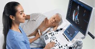
The procedure harnesses high-frequency sound waves (ultrasound) to generate a moving, active picture of your heart. A handheld device called a transducer sends sound waves safely through your chest wall. As these waves bounce back from your heart’s internal structures, they are instantly converted into vivid images on a clinical monitor, offering unparalleled insights into cardiac anatomy and motion.
Cardiologists rely on the 2D ECHO to effectively evaluate and manage a wide variety of conditions:
📋 How to Prepare
Preparing for this test is incredibly straightforward, as the process is completely painless. To ensure the most accurate results, simply follow these key steps:
- ✔ Clothing: Wear comfortable, loose-fitting clothes. You may be asked to remove your shirt during the test.
- ✔ Dietary Rules: Typically, no fasting is required unless you are also scheduled for a stress echocardiogram.
- ✔ Medications: Continue taking all prescribed medications unless your doctor specifically instructs otherwise.
- ✔ Communication: Always share any relevant medical history or current symptoms with the imaging technician.
⏱️ What Happens During the Test?
The entire procedure is quick, generally lasting only 15 to 30 minutes. While you might feel slight pressure from the wand, it is completely painless.
- Positioning: You will be asked to lie comfortably on an examination table, usually resting on your left side.
- Application of Gel: The technician applies a specialized, water-based gel to your chest to drastically improve sound wave transmission.
- Using the Transducer: The handheld device is gently moved across your chest to capture diverse angles of your heart.
- Image Capture: Real-time, moving images of your heart are fed directly to a monitor for the cardiologist to analyze.
Decoding Your Test Results
Interpreting a 2D ECHO requires the expert eye of a cardiologist. Should your test reveal any abnormalities, your doctor will discuss the severity and recommend targeted treatments or further testing.
Key Parameters Evaluated:
- Ejection Fraction (EF): Measures the exact percentage of blood pumped out with each beat. A healthy, normal EF ranges from 50% to 70%.
- Chamber Size & Wall Thickness: Checks for serious conditions like hypertrophy (thickened walls) or dilated cardiomyopathy.
- Valve Function: Confirms whether the heart valves are opening and closing tightly and effectively.
- Pericardial Effusion: Checks for any dangerous fluid buildup in the sac surrounding the heart.
🌟 What Constitutes a "Normal" Result?
- Proper heart chamber size and structural integrity.
- Smooth, normal valve function without any leaks or strict obstructions.
- Absolutely no fluid accumulation or visible abnormalities.
⊕ Key Advantages
⊖ Known Limitations
While highly effective, patients should be aware of a few constraints:
- • Operator Dependency: The final image quality can vary depending on the specific skill and experience of the technician.
- • Tissue Penetration: Sound waves may not provide perfectly detailed views in individuals who are heavily muscled or obese.
- • Supplementary Tests: Highly complex cardiac conditions might still require secondary imaging, such as a 3D ECHO, CT scan, or MRI.